Engineers Create Mini Microscope for Real-Time Brain Imaging
Researchers at the University of California, Davis, have created a miniaturized microscope for real-time, high-resolution and minimally invasive imaging of brain activity in mice. The device is a significant step toward revolutionizing how neuroscientists study the brain.
“What we are doing is creating technology to image brain activity in freely moving and behaving mice to open up the behavior paradigm,” said Weijian Yang, professor of electrical and computer engineering. “The goal is to create a device capable of enabling research into brain activity and behavior in mice in real time — to see how brain activity drives behavior or perception.”
The microscope will advance insights into how the brain works, which is expected to benefit human health by empowering the development of new and improved therapeutic strategies for brain disorders.
The first-of-its-kind imaging system, known as DeepInMiniscope, is described in a paper published Sept. 12 in Science Advances.
Iterative Design
DeepInMiniscope builds on Yang’s previous work to create a lensless camera capable of producing three-dimensional images from a single exposure.
Instead of a single bulky lens, Yang’s lensless camera used a thin mask hosting dozens of lenslets (minuscule lenses). Each lenslet captured a distinct perspective of the same object, after which computational algorithms synthesized the images to recreate an object in 3D.
This imaging system was well-suited for large objects in environments with minimal light scattering, such as robotic vision for part assembly, but struggled to capture the details of biological or biomedical samples. In living tissue, light scattering is prevalent, signal contrast tends to be low and reconstructing intricate features across a large volume of space is a computational problem.
DeepInMiniscope solves these issues with a new mask design containing more than 100 miniaturized, high-resolution lenslets and a novel neural network for image reconstruction.
The lenslets, together, form a microscope that can visualize brain tissue and brain activity without the need for deep-penetrating electrodes implanted by invasive surgery. However, the microscope is currently unable to see through opaque tissues like bone, making minimally invasive surgical procedures, like removing a small portion of the skull, a requirement.
"The device can be placed over the brain surface to image the activity inside rather than be inserted into the brain tissue," Yang said.
Yang's team is currently exploring methods to see through bone and remove the need for surgical intervention, aiming to make the microscope completely non-invasive.

Deep (Learning) Insights
DeepInMiniscope’s neural network combines principles from model-based iterative optimization and conventional deep neural networks.
Together, these methods create an unrolled neural network, where the architecture consists of multiple stages. Each stage functions as a mini-neural network that mimics a single iteration of a model-based optimization process. This design combines the strengths of both approaches: the robustness and interpretability of model-based iterative optimization, and the speed and efficiency of deep neural networks.
In the case of Yang’s microscope, the unrolled neural network allows for instantaneous, accurate and high-resolution reconstruction of fine details across a large 3D volume. It combines information from the 100 images taken by each lenslet into a single, coherent picture. Using this tool, Yang and his research team have recorded a mouse’s neuronal activity in real time.
“Our algorithm combines interpretability, efficiency, scalability and precision,” said Feng Tian, a postdoctoral researcher in Yang’s lab and first author on the corresponding paper. “It requires only a minimal amount of training data, yet it can robustly and accurately process large-scale datasets at high speed.”

Hat Trick
Essential to Yang’s aim of opening up neuroscience to real-time investigations of the behavior paradigm is to make his microscope small and ergonomic enough for a mouse to comfortably and safely wear as it moves freely.

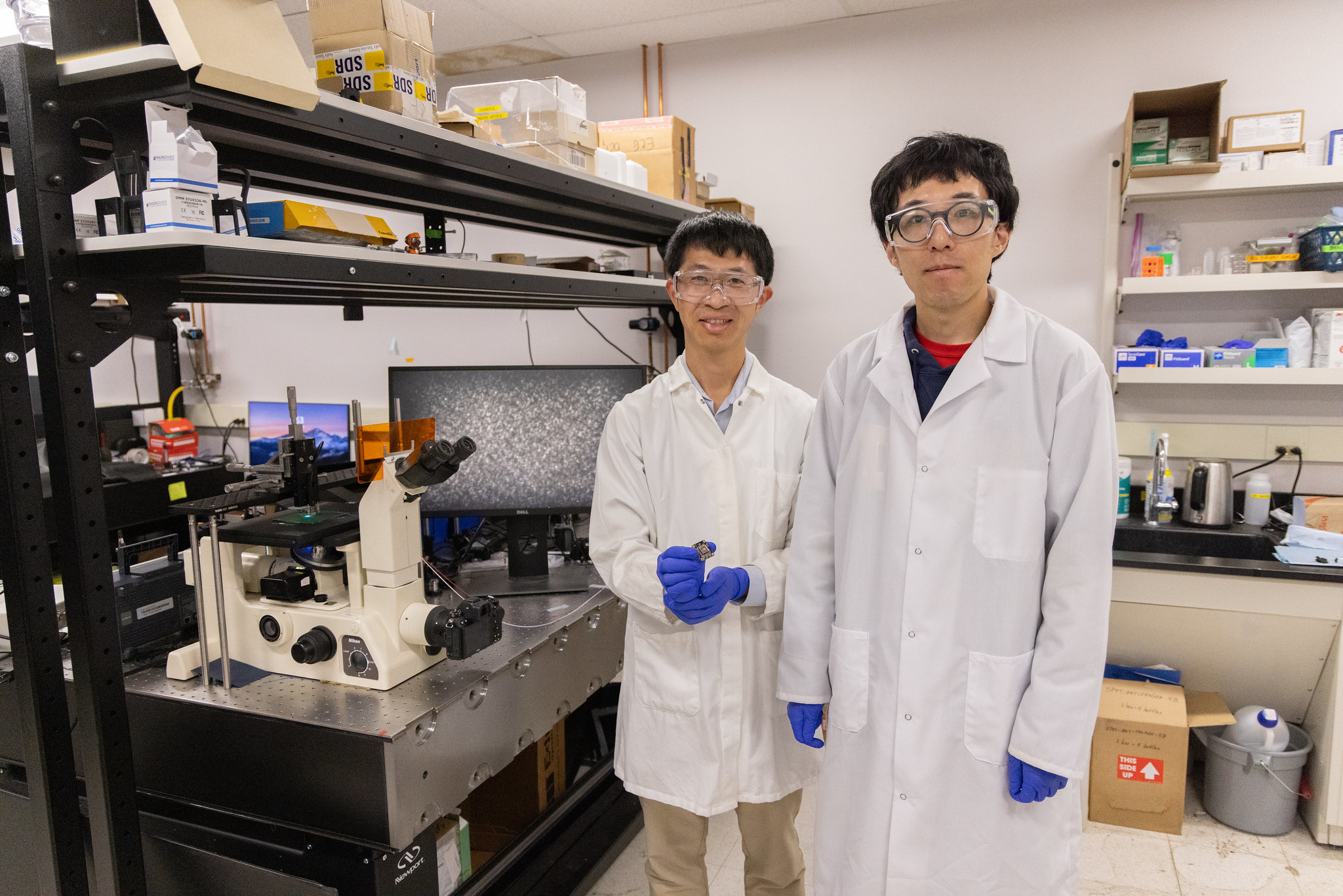
At just 3 square centimeters, about the size of a grape, and around the weight of four pennies at 10 grams, DeepInMiniscope is nearly there.
Where earlier, similar designs were constrained by the large footprint of a traditional camera, DeepInMiniscope uses a sensor as compact as a bare circuit board with an image sensor, rather than a self-contained and enclosed system.
Yang’s ultimate goal is a 2 square centimeter device, which he compares to the size of a hat for a mouse. Additionally, for the next iteration, Yang wants to make the device cordless.
“By enabling real-time observation of brain activity in freely behaving mice, this technology not only advances our fundamental understanding of how the brain processes information and drives behavior, but also contributes to improving our understanding of brain disorders and the development of future therapeutic strategies in humans.”
Media Resources
- Weijian Yang, Electrical and Computer Engineering, [email protected]
- Matt Marcure, College of Engineering, [email protected]